Can't intubate, can't oxygenate (CICO) — emergency front-of-neck access
Airway emergency · Perioperative & Anaesthesia
Physician-written · Verified by three sets of doctors · Reviewed November 2026
Bedside summary
CICO is the failure of all upper-airway oxygenation methods (face mask, supraglottic airway and tracheal tube) with desaturation — a true time-critical emergency requiring immediate surgical front-of-neck access (eFONA) to prevent hypoxic brain injury and death.
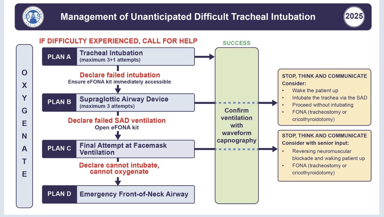
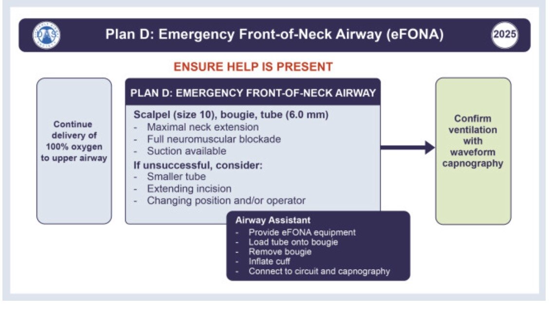
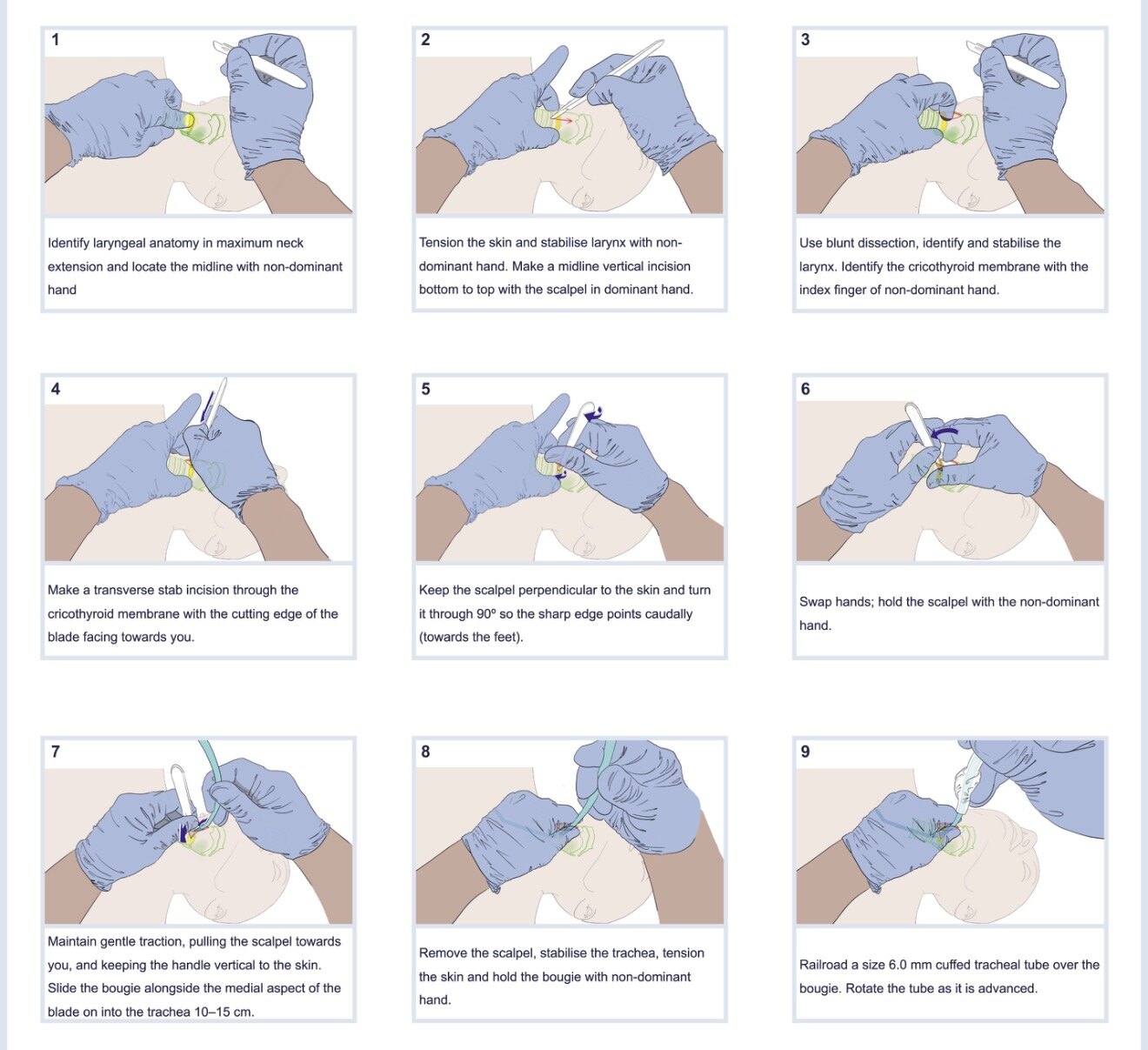
Figures & algorithms
Red flags
- SpO2 falling despite optimised mask/SGA/intubation attempts
- Absent capnography trace with no chest rise
- Bradycardia heralding hypoxic cardiac arrest
- Impalpable neck landmarks (obesity, haematoma, radiotherapy) delaying access
- Repeated laryngoscopy attempts wasting time instead of committing to FONA
Assessment
Differential diagnosis
- Failed intubation with failed SGA and failed mask ventilation
- Laryngospasm progressing to complete obstruction
- Airway tumour / supraglottic pathology / angioedema
- Foreign body or blood/soiling obscuring the glottis
- Severe laryngeal trauma or distorted anatomy
- Massive airway oedema (burns, anaphylaxis, radiotherapy)
Investigations
- Continuous SpO2 and waveform capnography (absent trace confirms no ventilation)
- Visual/clinical assessment of chest rise and face-mask seal
- Palpation of cricothyroid membrane / neck landmarks
- Direct or video laryngoscopic view grade (Cormack–Lehane)
- Post-event ABG and chest X-ray to assess oxygenation/tube position
- Fibreoptic/flexible scope confirmation of tube position after rescue
Management
- Declare CICO aloud and call for help, difficult airway trolley and a second anaesthetist/surgeon
- Give 100% oxygen and make a final optimised attempt at mask ventilation (2-person, adjuncts, full relaxation) and one best-attempt SGA if not already tried
- Ensure full neuromuscular blockade — give/repeat suxamethonium or rocuronium before cutting
- Commit early to scalpel-bougie-tube cricothyroidotomy; do not delay for repeated laryngoscopy
- Extend the neck, stabilise the larynx with non-dominant hand, identify the cricothyroid membrane
- Palpable anatomy: transverse stab incision through the membrane, rotate blade 90°, keep blade in situ
- Impalpable anatomy: 8–10 cm vertical midline incision, finger dissection to larynx, then transverse stab
- Slide bougie into the trachea alongside the scalpel (angle caudally), confirm tracheal clicks/hold-up
- Railroad a size 6.0 cuffed tube over the bougie, remove bougie, inflate cuff
- Confirm placement with waveform capnography; ventilate gently and secure the tube
- Hand-ventilate, reassess oxygenation and summon ENT/surgical airway support
- Document, debrief, arrange critical-care admission and airway follow-up
Drugs & doses
Suxamethonium 1–1.5 mg/kg IV (or 4 mg/kg IM, max 200 mg, if no IV access)
Rocuronium 1.2 mg/kg IV for full relaxation if suxamethonium contraindicated
Sugammadex 16 mg/kg IV only if reversing rocuronium is a genuine rescue option (does NOT help once CICO declared — proceed to FONA)
Oxygen 100% / 15 L/min via best available device throughout
Lidocaine 1% infiltration only if time permits and patient not fully anaesthetised (usually not applicable in emergency)
Disposition
Immediate ICU admission after securing the airway; involve ENT/head-and-neck surgery urgently, keep the FONA tube until a definitive airway plan and formal airway review are completed.
Complications & pitfalls
- Hypoxic brain injury or death from delayed access
- Haemorrhage from cricothyroid/thyroid vessels
- Tube malposition (pretracheal, oesophageal) or barotrauma
- Posterior tracheal wall / oesophageal perforation
- Surgical emphysema, pneumothorax, later subglottic stenosis
Exam pearls
- 'Scalpel-finger-bougie' — the finger maintains the anatomical path once the airway is incised
- Full neuromuscular blockade improves conditions for both ventilation and FONA — do not withhold relaxant
- Human factors kill: a fixation on intubation delays the life-saving cut; verbalise the transition
- Use a size 10 scalpel, bougie and 6.0 cuffed tube — a standardised single technique saves cognitive load
- Practise the technique regularly; the first time should never be the real thing
Disclaimer: Educational decision support for qualified clinicians and students — not a substitute for clinical judgement, local protocols or a full drug reference. Confirm every dose and contraindication against your formulary before prescribing.