Acute optic neuritis
Inflammatory · Neurology
Physician-written · Verified by three sets of doctors · Reviewed November 2026
Bedside summary
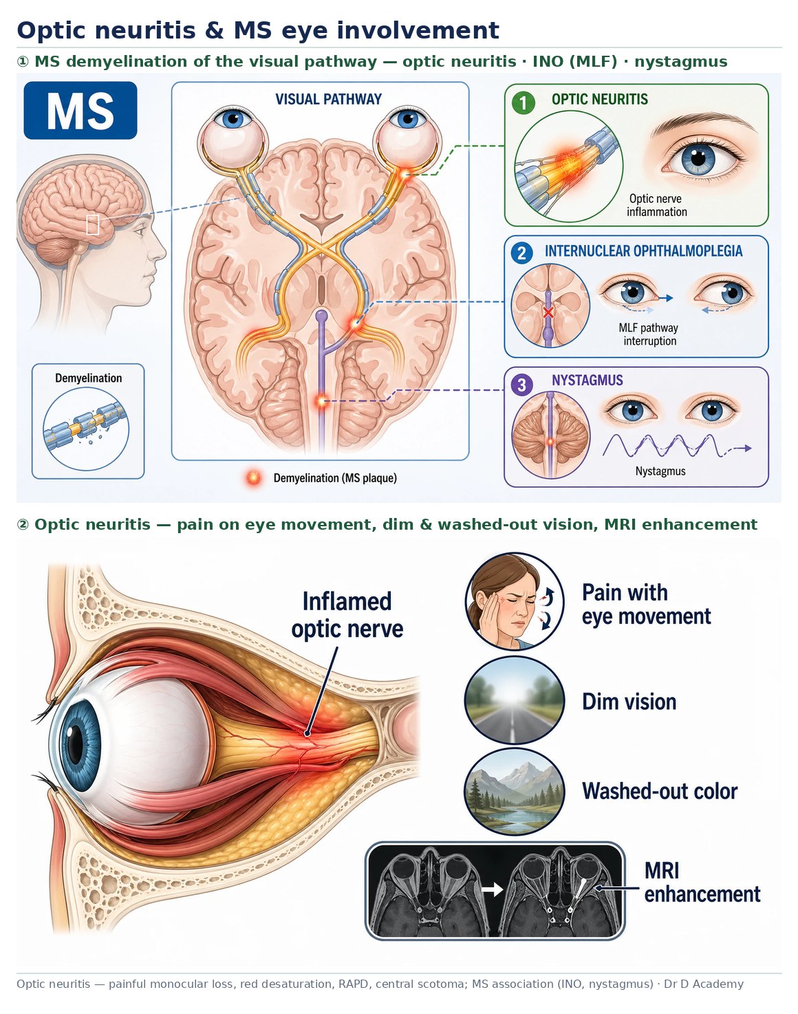
Subacute, usually unilateral visual loss with pain on eye movement, reduced colour vision and a relative afferent pupillary defect. Often the presenting event of multiple sclerosis, but atypical features suggest NMOSD or MOG-associated disease.
Figures & algorithms
Red flags
- Bilateral simultaneous or severe visual loss (suggests NMOSD/MOG)
- No pain, or progressive painless loss (suggests ischaemic/compressive cause)
- Poor recovery beyond expected timeframe
Assessment
Differential diagnosis
- Anterior ischaemic optic neuropathy
- Neuromyelitis optica spectrum disorder
- MOG antibody-associated disease
- Leber hereditary optic neuropathy
- Compressive or infiltrative optic neuropathy
Investigations
- Visual acuity, colour vision and relative afferent pupillary defect testing
- MRI brain and orbits with contrast (and spine if NMOSD suspected)
- Aquaporin-4 and MOG antibodies if atypical/severe/bilateral
- Fundoscopy (often normal in retrobulbar neuritis)
- Bloods and CSF if inflammatory/infective cause considered
Management
- Confirm optic neuritis clinically (pain on movement, RAPD, dyschromatopsia)
- Arrange MRI brain and orbits to assess demyelination and exclude compression
- Send NMOSD/MOG antibodies if atypical, bilateral, severe or poorly recovering
- Discuss high-dose corticosteroid to speed recovery (does not change final acuity in typical MS-related cases)
- Refer to neurology/ophthalmology for follow-up and disease-modifying decisions
Drugs & doses
High-dose IV or oral methylprednisolone to hasten recovery
Avoid low-dose oral prednisolone alone (associated with higher recurrence in classic trials)
Disease-modifying therapy decisions by neurology
Disposition
Urgent neurology/ophthalmology referral; steroid decision individualised.
Complications & pitfalls
- Permanent visual deficit (higher in NMOSD)
- Conversion to multiple sclerosis
- Recurrence
Exam pearls
- Painless progressive loss over weeks is not typical optic neuritis - reconsider compression or ischaemia
- Atypical or severe cases need aquaporin-4 and MOG testing before labelling as MS
Disclaimer: Educational decision support for qualified clinicians and students — not a substitute for clinical judgement, local protocols or a full drug reference. Confirm every dose and contraindication against your formulary before prescribing.