Superior vena cava (SVC) syndrome
Protocol · Vascular
Physician-written · Verified by three sets of doctors · Reviewed November 2026
Bedside summary
Obstruction of the SVC — face/neck/arm swelling and head fullness (worse bending/lying), distended neck + chest-wall veins, facial plethora, dyspnoea. Malignancy (lung cancer ~50%, SCLC, NHL) causes 60–85% (the first sign of an undiagnosed tumour in up to 60%); catheter/device thrombosis now causes 20–40%. True emergencies = stridor/laryngeal oedema or cerebral oedema (headache, confusion, coma).
Figures & algorithms
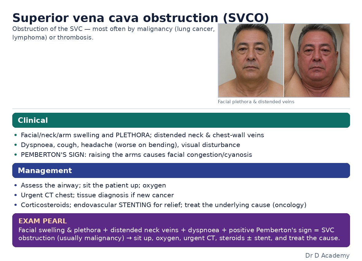
Assessment
Investigations
- Contrast-enhanced CT chest (with upper-limb contrast) — the initial test; MR venography if iodine allergy
- Get a TISSUE diagnosis before tumour-directed therapy (60% have no known cancer) — UNLESS a true airway/CNS emergency
- Grade severity by airway/cerebral features — this drives urgency
Management
- Supportive: head up + oxygen; avoid upper-limb IV lines/IM injections; cautious diuresis
- True emergency (airway/cerebral oedema) → stabilise ABC then URGENT endovascular stenting ± thrombectomy (success 95–100%, rapid relief); empiric radiotherapy is no longer first-line (obscures histology)
- Otherwise treat the tumour — chemotherapy for chemosensitive (SCLC/NHL/germ cell), radiotherapy for others; anticoagulate catheter/thrombus-related disease
- Steroids only for a steroid-responsive tumour (lymphoma/thymoma — not before histology) or to bridge emergency RT
Disposition
Admit + oncology. Life-threatening airway/cerebral features → urgent interventional radiology for stenting.
Disclaimer: Educational decision support for qualified clinicians and students — not a substitute for clinical judgement, local protocols or a full drug reference. Confirm every dose and contraindication against your formulary before prescribing.