Central retinal artery occlusion
Sight-threatening · Ophthalmology
Physician-written · Verified by three sets of doctors · Reviewed November 2026
Bedside summary
Sudden, painless, profound monocular visual loss — an ocular “stroke”. Retina tolerates ischaemia for only ~90 min before irreversible loss.
Figures & algorithms
Red flags
- Jaw claudication, scalp tenderness, raised ESR (GCA)
- Other focal neurology (stroke)
Assessment
Differential diagnosis
- Central retinal vein occlusion
- Giant cell arteritis (AION)
- Vitreous haemorrhage
- Retinal detachment
- Stroke
Investigations
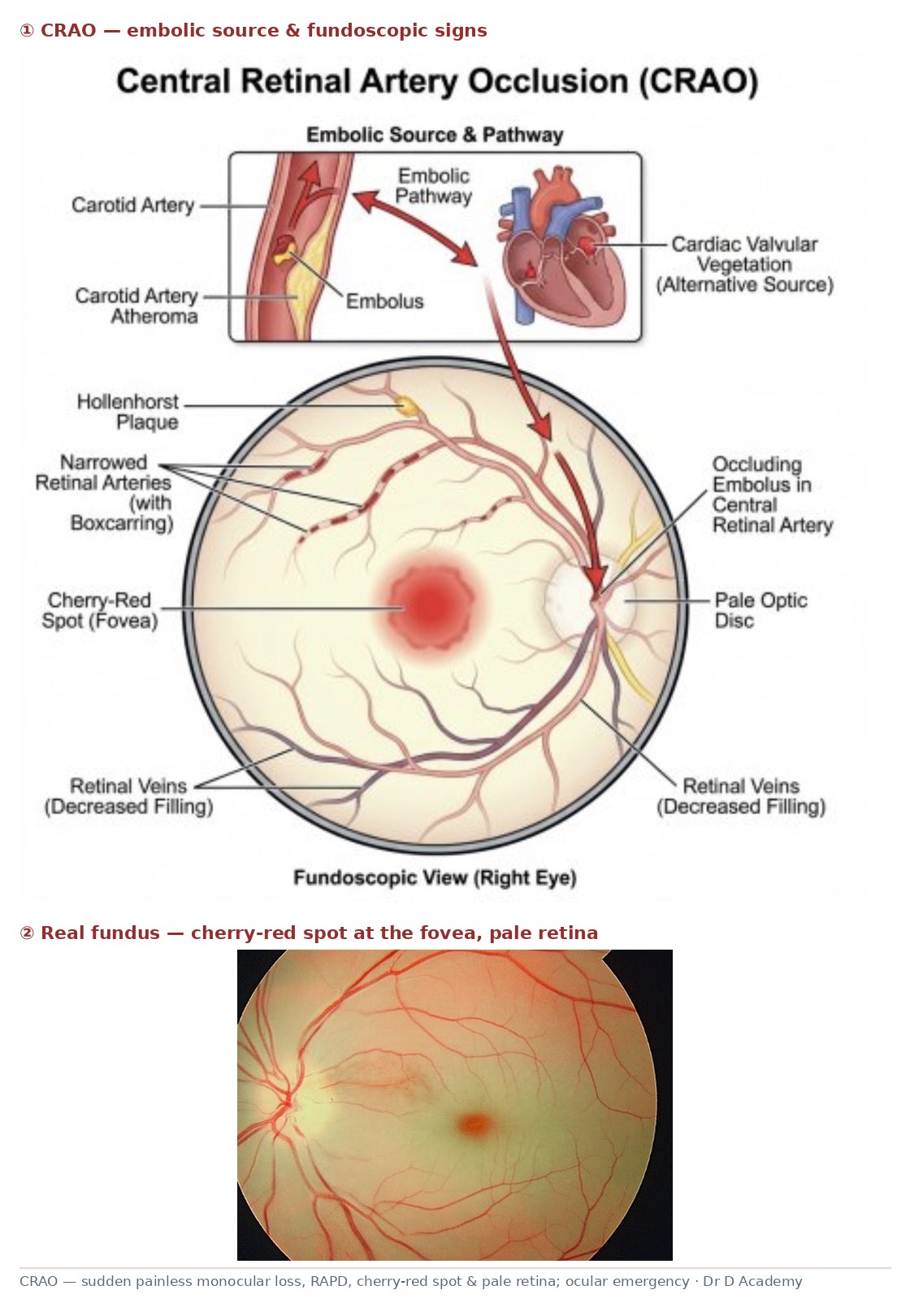
- Visual acuity; RAPD present; fundoscopy (pale retina, cherry-red spot)
- ESR/CRP urgently (exclude giant cell arteritis)
- Treat as a stroke equivalent — vascular workup
Management
- Immediate ophthalmology + stroke pathway referral
- Attempt measures within the window: ocular massage, lower IOP (acetazolamide), rebreathing
- Exclude and treat giant cell arteritis (high-dose steroids if suspected)
- Investigate embolic source (carotid, cardiac); manage vascular risk
- Counsel on guarded prognosis
Drugs & doses
Acetazolamide; ocular massage
High-dose steroids if GCA suspected
Disposition
Emergency ophthalmology + stroke/TIA pathway.
Complications & pitfalls
- Permanent blindness
- Underlying stroke/vascular disease
Exam pearls
- Treat as a stroke — time-critical, full vascular workup
- Always exclude giant cell arteritis (steroids save the other eye)
- Cherry-red spot with a pale retina is classic
Act immediately (massage/lower IOP), exclude GCA, and treat as an ocular stroke with a vascular workup.
Disclaimer: Educational decision support for qualified clinicians and students — not a substitute for clinical judgement, local protocols or a full drug reference. Confirm every dose and contraindication against your formulary before prescribing.